Tagged: Tumors

Novel GEM Model Unveils PLK1’s Role in Tumorigenesis

In a new editorial, researchers discuss a study using their team’s new genetically engineered mouse (GEM) model to assess PLK1 as a driver of oncogenic transformation.

Figure 1: The role of PLK1 in tumorigenesis and cancer heterogeneity. GEM Model

On the bright side, polo-like kinase 1 (PLK1) is considered a master regulator of the ever-important cell cycle. On the dark side, PLK1 expression (at both the mRNA and protein level) has shown to be upregulated in tumor cells, suggesting that PLK1 may also contribute to tumorigenesis. Despite this direct association, researchers studying the role of PLK1 in cancer have encountered a problem: a lack of appropriate animal models for experimentation.

“Even though studies have suggested that PLK1 contributes to tumorigenesis, the ability of PLK1 to drive oncogenic transformation on its own in vivo was still questionable due to a lack of sophisticated animal models for experimentation [18, 19].”

This problem may have been solved in 2021. In a new editorial paper, researchers Lilia Gheghiani and Zheng Fu from Virginia Commonwealth University discuss a recent study using their team’s new genetically engineered mouse (GEM) model to assess the ability of PLK1 to be a sole driver of oncogenic transformation in vivo. Their editorial was published in Oncotarget’s Volume 14 on June 27, 2023, and entitled, “The dark side of PLK1: Implications for cancer and genomic instability.”

PLK1 in Tumorigenesis

“To address this important scientific question, we generated a new genetically engineered mouse (GEM) model using the CAGGS (cytomegalovirus (CMV) early enhancer/chicken β-actin) promoter to drive exogenous PLK1 expression, allowing its ubiquitous and robust gene expression in transgenic mice [20].”

In an effort to determine if PLK1 overexpression causes tumors, the researchers created a new GEM mouse model that expresses high levels of PLK1. These high levels caused various types of spontaneous tumors. The increased PLK1 levels caused defects in cell division and resulted in abnormal numbers of centrosomes and compromised cell cycle checkpoints. This allowed for the accumulation of chromosomal instability, leading to abnormal numbers of chromosomes and tumor formation. In human cancers, higher PLK1 expression was associated with an increase in genome-wide copy number alterations. Their study provides evidence that abnormal PLK1 expression can trigger chromosomal instability and tumor formation, suggesting potential therapeutic opportunities for cancers with chromosomal instability.

“In summary, this study provides a novel GEM model that recapitulates the increased PLK1 expression observed in many human cancers and demonstrates that PLK1 overexpression drives spontaneous tumor formation in multiples organs in mouse, revealing the dark side of PLK1 as a potent proto-oncogene.”

Conclusions

In conclusion, the limitations of previous studies on PLK1 and its role in cancer have been partially addressed by the development of the new GEM model created by these researchers. This model allowed the team to examine PLK1’s ability to drive oncogenic transformation in vivo. Their study demonstrates that overexpression of PLK1 leads to the formation of spontaneous tumors in multiple organs, highlighting the dark side of PLK1 as a potent proto-oncogene. The findings of this study provide valuable insights into the role of PLK1 in tumorigenesis and suggest potential therapeutic opportunities for cancers associated with chromosomal instability. This breakthrough in animal models opens up new avenues for further research in understanding the mechanisms underlying PLK1-related tumorigenesis and developing targeted therapies to combat cancer.

“Alternative therapeutic strategies, such as co-delivery systems using nanoparticles or combination therapies, are under development in order to enhance the efficacy of PLK1 inhibition [2528]. With expanding discoveries of PLK1 function and mechanisms of action, we hope that PLK1-targeted therapies will soon join the frontlines in the fight against cancer.”

Click here to read the full editorial paper in Oncotarget.

ONCOTARGET VIDEOS: YouTube | LabTube | Oncotarget.com

Oncotarget is an open-access, peer-reviewed journal that has published primarily oncology-focused research papers since 2010. These papers are available to readers (at no cost and free of subscription barriers) in a continuous publishing format at Oncotarget.com. Oncotarget is indexed/archived on MEDLINE / PMC / PubMed.

Click here to subscribe to Oncotarget publication updates.

For media inquiries, please contact media@impactjournals.com.

Trending with Impact: Benign, Borderline, and Malignant Breast Tumors


Researchers conducted a 2021 study to better characterize phyllodes tumors and other breast fibroepithelial lesions in order to improve diagnosis and treatment for patients.

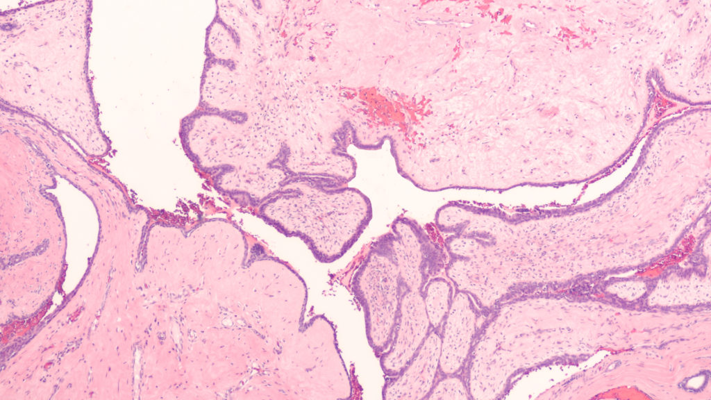
Photomicrograph showing histology of a benign phyllodes tumor of the breast, from sections of an excision specimen (lumpectomy).

The Trending with Impact series highlights Oncotarget publications attracting higher visibility among readers around the world online, in the news, and on social media—beyond normal readership levels. Look for future science news and articles about the latest trending publications here, and at Oncotarget.com.

Listen to an audio version of this article

Thankfully, around 80% of lumps found in human breasts turn out to be benign, or indolent, fibroadenoma (FAD). FADs fall into a category of breast fibroepithelial lesions (FELs), which include many heterogeneous pathological tumors, ranging from benign FADs to rare and potentially aggressive phyllodes tumors (PTs). After examination by a physician, these FELs may be diagnosed as either benign, borderline, or malignant. 

However, there is a need to improve accurate diagnosis and distinction between FELs using a marker-based diagnostic approach. In an effort to better characterize FELs, researchers from India’s CSIR-Centre for Cellular and Molecular Biology, Institute of Bioinformatics, Gandhi Hospital, Government Medical College, and Manipal Academy of Higher Education conducted a trending 2021 study, titled: “Quantitative proteome profiling stratifies fibroepithelial lesions of the breast.”

“The current grading system remains unreliable in differentiating these tumors due to histological heterogeneity and lack of appropriate markers to monitor the sudden and unpredictable malignant transformation of PTs.”

The Study

To begin identifying the differentially expressed genes and proteins among FADs and PTs in benign, borderline, and malignant states, the researchers conducted quantitative global proteomics on Formalin-Fixed Paraffin-Embedded (FFPE) tissue sections. They conducted a principal component analysis of the protein expression matrix to identify the overlapping proteomic profiles among FELs.

“Interestingly, we observed FADs and benign PTs clustered together compared to borderline and malignant ones, albeit with overlapping protein expression profiles.”

When FADs were compared with benign PTs, the researchers identified 32 proteins in FAD that were differentially regulated. The researchers elucidated many important distinctions between benign, borderline, and malignant FADs and PTs, and identified at least three potential prognostic markers that may aid in patient diagnosis and treatment. The progression of PTs from borderline to malignant and their mechanistic framework was clearly explained by the researchers in this study.

“The presence of extensive ECM proteins and EMT markers led us to hypothesize a model of deposition and degradation of these proteins thus triggering ECM remodeling and EMT acquisition in borderline PTs leading to its malignant state. Enrichment of platelet degranulation factors in malignant PT indicates active angiogenesis during this transformation.” 

The Study

“Herein, our initial findings suggest that MUCL1, HTRA1, and VEGFD can be used as potential proteomic markers that could augment existing diagnosis, and help in monitoring the progression of the disease.” 

Additional characterization of FELs using different omics platforms was recommended by the researchers to help better understand and manage the dynamics of PTs and malignant breast tumors.

“The present work shed light on a brief mechanistic framework of PTs aggressive nature and present potential biomarkers to differentiate overlapping FELs that would be of practical utility in augmenting existing diagnosis and disease management for this rare tumor.”

Click here to read the full scientific study, published in Oncotarget.

Oncotarget is a unique platform designed to house scientific studies in a journal format that is available for anyone to read—without a paywall making access more difficult. This means information that has the potential to benefit our societies from the inside out can be shared with friends, neighbors, colleagues, and other researchers, far and wide.

For media inquiries, please contact media@impactjournals.com.