Tagged: Mouse Model

Melatonin in Cancer Therapy: Lessons From 50 Years of Research

In a new research perspective, researchers discuss melatonin’s effects on cancer and the key importance of the timing of administration.

Melatonin in Cancer Therapy: Lessons From 50 Years of Research

In the realm of cancer research, the potential of melatonin as an anti-cancer agent has garnered significant attention. Over the past 50 years, numerous studies have been conducted to investigate the effects of melatonin on tumor growth and development in mice. These studies have provided valuable insights into the complex relationship between melatonin and carcinogenesis.

In a new research perspective, researchers Vladimir N. Anisimov and Alexey G. Golubev from N.N. Petrov National Medical Research Center of Oncology wrote about the history of studies of melatonin effects on cancer in mice. Their paper was published in Oncotarget on December 12, 2023, entitled, “Melatonin and carcinogenesis in mice: the 50th anniversary of relationships.”

Early Discoveries and Controversies

In 1973, Vladimir N. Anisimov and his coauthors made a groundbreaking discovery by demonstrating the inhibitory effect of melatonin on transplantable mammary tumors in mice. This pivotal study laid the foundation for subsequent investigations into the potential anti-cancer properties of melatonin. However, early studies encountered controversies regarding the consistency of melatonin’s effects on in vivo cancer models. The lack of consistency in these studies prompted further exploration of the factors influencing melatonin’s efficacy.

Importance of Timing in Melatonin Administration

One of the crucial findings in melatonin research is the significant impact of timing in melatonin administration. Bartsch and Bartsch demonstrated that the effects of melatonin on cancer in mice depend on the time of treatment. The administration of melatonin in the morning stimulated tumor growth, while late afternoon administration inhibited it. This observation highlighted the importance of considering animal conditions and the systemic effects of melatonin when evaluating its anti-cancer properties. These systemic effects may not be evident in cell cultures or ex vivo explants.

Murine Models for Melatonin and Cancer Studies

Murine models have played a pivotal role in elucidating the effects of melatonin on various types of cancer. These models have provided valuable insights into the potential utility of melatonin in oncology. Some of the notable murine models include mice grafted with murine tumors, chemically induced tumors, spontaneous carcinogenesis in mice, transgenic HER2/neu oncogene-bearing mice, and nude mice grafted with human prostate tumors. These models have allowed researchers to evaluate not only the effects of melatonin on cancer development but also its impact on the efficacy and side effects of anticancer therapies.

Melatonin’s Effects on Spontaneous Tumor Incidence

One intriguing finding in murine studies is the effect of melatonin on spontaneous tumor incidence. Anisimov et al. showed that lifelong treatment of mice with melatonin decreased the incidence of spontaneous tumors, particularly mammary carcinomas, but only at a low concentration of melatonin in drinking water. Interestingly, this effect was not observed at a high melatonin concentration. These findings suggest that the dose of melatonin may play a crucial role in its anti-cancer effects.

Melatonin’s Role in Potentiating Cytotoxic Therapy

Another area of interest in melatonin research is its potential to enhance the efficacy of cytotoxic therapy against tumors. Panchenko et al. demonstrated that the timing of melatonin administration relative to cytotoxic drug administration significantly influenced its potentiating effect on cytotoxic therapy in HER2/neu transgenic mice. This finding highlights the importance of optimizing the timing of melatonin administration in combination with other cancer treatments.

Melatonin’s Protective Effects on Side Effects

Beyond its direct anti-cancer effects, melatonin has shown promise in alleviating the side effects of cytotoxic drugs and radiation therapy. Several murine models have demonstrated the ability of melatonin to mitigate the side effects associated with these treatments. For example, melatonin was shown to alleviate the depression syndrome in mice treated with the alkylating agent temozolomide used in brain cancer therapy. Additionally, melatonin has been found to protect against ovarian follicle depletion caused by cisplatin, a commonly used chemotherapy drug. These findings suggest that melatonin may have a broader role in cancer treatment by reducing the adverse effects of traditional therapies.

Melatonin’s Effects on Metastasis and Epithelial-Mesenchymal Transition

Metastasis is a significant challenge in cancer treatment, and melatonin has shown promise in inhibiting metastatic spread. In nude mice grafted with human gastric cancer, melatonin was found to suppress lung metastases development by inhibiting the epithelial-to-mesenchymal transition (EMT). The inhibition of EMT by melatonin has also been observed in other murine models, highlighting its potential as an anti-metastatic agent. Given the crucial role of EMT in primary cancer and metastasis development, these findings have significant implications for oncology research.

Melatonin and Inflammation

Chronic inflammation is increasingly recognized as a contributing factor in cancer development and progression. Melatonin has been found to modulate inflammatory processes in murine models. In a murine model of low-grade inflammation, melatonin inhibited EMT), suggesting a potential role in suppressing cancer-related inflammation. While the direct anti-inflammatory effects of melatonin require further investigation, these findings shed light on the multifaceted mechanisms through which melatonin may exert its anti-cancer effects.

Clinical Applications and Promising Results

The employment of melatonin in clinical settings beyond its established fields does not require licensing, making it more readily accessible for testing novel applications in cancer treatment. Promising clinical results have already been reported, such as increased overall survival in prostate cancer patients with poor prognosis after combined hormone radiation treatment. These findings highlight the potential translational impact of murine studies and underscore the importance of continued research to fully understand the clinical implications of melatonin in cancer therapy.

Conclusion

Over the past 50 years, murine models have provided valuable insights into the relationship between melatonin and carcinogenesis. These studies have shed light on the importance of timing in melatonin administration, its effects on tumor incidence and metastasis, as well as its role in potentiating cytotoxic therapy and mitigating side effects. While the precise mechanisms underlying melatonin’s anti-cancer effects require further exploration, the promising results observed in both preclinical and clinical studies warrant continued investigation. As researchers continue to unravel the complexities of melatonin’s interactions with cancer, new opportunities for therapeutic interventions may emerge, offering hope for improved cancer treatment outcomes.

“The […] main lesson being that the systemic in vivo effects of melatonin on animals may overwhelm the in vitro effects found using tissue explants or cell cultures. In particular, the timing of melatonin administration is of crucial importance for using the drug, which is freely available over [the] counter and thus needs no licensing for its applications in oncology.”

Click here to read the full research perspective in Oncotarget.

Oncotarget is an open-access, peer-reviewed journal that has published primarily oncology-focused research papers since 2010. These papers are available to readers (at no cost and free of subscription barriers) in a continuous publishing format at Oncotarget.com. Oncotarget is indexed/archived on MEDLINE / PMC / PubMed.

Click here to subscribe to Oncotarget publication updates.

For media inquiries, please contact media@impactjournals.com.

Trending with Impact: Murine Model of Cancer-derived Myocardial Damage

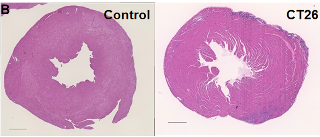
Researchers in this study employed one of the few available murine cachexia models and validated its ability to be used in future studies of cancer-derived myocardial damage.

Part of Figure 2: Alterations in the myocardium of CT26-inoculated BALB/c mice.

The Trending with Impact series highlights Oncotarget publications attracting higher visibility among readers around the world online, in the news, and on social media—beyond normal readership levels. Look for future science news and articles about the latest trending publications here, and at Oncotarget.com.

Listen to an audio version of this article

Cachexia, a complex metabolic syndrome characterized in part by the loss of muscle mass, can account for up to 30% of all cancer-related deaths. Myocardial atrophy, or cardiac remodeling/degradation, is a phenotype of cachexia and a common cause of death.  

“The causes of cancer-derived myocardial impairment might be the effects of cancer itself, background heart disease, and influence of cancer treatments; however, they have not been given much clinical importance, and specific treatment efforts are delayed [8].”

Researchers from Nara Medical University, Hanna Central Hospital, and Hoshida Minami Hospital in Nara and Osaka, Japan, and Nantong University in Jiangsu Province, China, note that while myocardial damage in cancer patients is known to be a cause of death, there are few murine cachexia models available to evaluate cancer-derived heart disorders. Thus, there is a need for further studies that may allow researchers to establish an intervention to prevent myocardial damage in cancer patients.

“In this study, we used the mouse cancer cachexia model that we previously established [14] to examine the status of cancer-derived myocardial impairment reported in literature, and validate our model for studying cancer-derived myocardial impairment.”

The Study

Some causes of cancer-derived myocardial impairment have been reported as cancer-induced cytokines, oxidative stress, depletion of antioxidants, and protein catabolism as a result of AKT/mTOR inhibition.

“Despite these advances in our understanding, the multifactorial mechanisms underlying cancer-derived myocardial impairment remain incompletely understood, necessitating further investigations to elucidate the molecular mechanisms and prevent myocardial damage in cancer patients.”

The researchers previously established a mouse cancer cachexia model. In this study, they aimed to validate their model by employing it in the examination of cancer-derived myocardial impairment that has been reported in previous literature. Their study enlisted the mouse model, CT26 colon cancer cell cultures, protein extraction, histological analysis, immunoblot analysis, enzyme-linked immunosorbent assay (ELISA), mitochondrial stress tests (Seahorse assay), glycolytic stress tests, and statistical analysis. 

Conclusion

“In summary, our established mouse cachexia model showed various myocardial changes associated with cancer cachexia such as oxidative stress in the myocardium, energy metabolism, autophagy, and inflammatory cytokines.”

Results obtained by the researchers in this study using their mouse cachexia model are congruent with previously reported results about cancerous myocardial damage, and therefore provide reasonable evidence that it may be used in future studies.

“The established mouse cachexia model can therefore be considered useful for analyzing cancer-derived myocardial damage.”

Click here to read the full scientific study, published in Oncotarget.

Oncotarget is a unique platform designed to house scientific studies in a journal format that is available for anyone to read—without a paywall making access more difficult. This means information that has the potential to benefit our societies from the inside out can be shared with friends, neighbors, colleagues and other researchers, far and wide.

For media inquiries, please contact media@impactjournals.com.
Trending with Impact: Bacterial Therapy Experiments in Prostate Cancer

Researchers reveal their positive findings from a study of bacterial cancer therapy using a strain of Salmonella typhimurium in mouse-modeled prostate cancer.

PC-3 human prostate cancer cells stained with blue Coomassie, under a differential interference contrast microscope. - Image
PC-3 human prostate cancer cells stained with blue Coomassie, under a differential interference contrast microscope.

The Trending with Impact series highlights Oncotarget publications attracting higher visibility among readers around the world online, in the news, and on social media—beyond normal readership levels. Look for future science news and articles about the latest trending publications here, and at Oncotarget.com.

Listen to an audio version of this article

Over the past few decades, numerous studies have emerged using the promising strategy of bacteria as vehicles to deliver drugs or genes in tumor‐targeted therapies. Researchers say that bacterial cancer therapy may be able to overcome some of the limitations that conventional cancer therapy is stunted by, including the development of drug resistance. 

Researchers in this study—from Yale University in Connecticut, the University of Missouri, the Harry S. Truman Memorial Veterans Hospital, and the Cancer Research Center in Missouri, and DeSales University in Pennsylvania, U.S.—used a Salmonella typhimurium strain (CRC2631) of bacteria (previously reported to have tumor-targeting capabilities) in prostate cancer-positive mouse-models and evaluated its toxicity, targeting ability, and genetic stability.

“Here, we report the toxicological and in vivo tumor-targeting profiles of CRC2631 in the syngeneic and autochthonous mouse model of aggressive prostate cancer, TRAMP (Transgenic Adenocarcinoma of Mouse Prostate).”

“The B6FVB TRAMP model recapitulates some of the key genetic aspects of human prostate cancer.”

The Study

“VNP20009 is considered as the safety benchmark in bacterial cancer therapy development because it has been safely administered in human cancer patients [7, 30].”

“To determine the safety profile of CRC2631, we performed CRC2631 and VNP20009 comparative toxicological studies in TRAMP animals.”

The team focused on measuring toxicity through treatment-related weight loss and lethality. Groups of 14-week-old B6FVB TRAMP-positive mice were scanned using magnetic resonance imaging. Four mice were dosed with CRC2631, and four were dosed with VNP20009; both treated four times per week. Mice were weighed and monitored daily for four weeks.

Since the CRC2631 bacteria are cleared out through the liver, the researchers also sought to establish the impact of CRC2631 on liver pathology in this bacterial cancer therapy. Two groups of 31-week-old B6FVB TRAMP-positive mice were observed, one treated with four doses of CRC2631 and the other with saline (the control group) at three-day intervals. They used histological staining in the liver to observe differences in necrosis, inflammation, and extramedullary hematopoiesis between CRC2631 and the control group. The team then tested for lethality and the maximum tolerated dose of CRC2631.

“Next, we sought to determine the in vivo tumor-targeting capability of CRC2631 in TRAMP animals.”

Using fluorescence imaging and a chloramphenicol resistance cassette, researchers were able to observe the biodistribution of CRC2631 to determine its tumor-targeting capability in TRAMP-positive mice. Since they knew that CRC2631 is filtered through the liver and that enriched colonies may be found here, researchers used the liver as a way to compare the bacterial load in tumor tissues.

The researchers also tested CRC2631’s genetic stability by gauging its likelihood of regaining toxicity and/or losing tumor targeting capability by performing longitudinal, whole genome sequencing and short nucleotide polymorphism analyses.

“To determine the genetic stability of CRC2631 inside the host, we performed longitudinal whole genome sequencing and short nucleotide polymorphism (SNP) analyses of CRC2631 prior to treatment and tumor-passaged CRC2631 in B6 TRAMP (+) mice.”

In vitro, CRC2631 directly kills prostate cancer cells, however, in vivo, it does not lead to decreased tumor burden. The researchers believe this may be due to the effects of some kind of resistance mechanism in vivo, and tested a combined treatment method of CRC2631 and Invivomab—a checkpoint blockade—in the mouse model.

“CRC2631 targets and directly kills murine and human prostate cancer cells in vitro (Supplementary Figure 2), raising the possibility that unknown resistance mechanisms protect tumor cells from CRC2631-mediated cell death in vivo.”

Results

Researchers explain that in the first two weeks of the study, mice treated with CRC2631 and VNP20009 lost a comparable amount of weight. However, in the second half of the study, VNP20009-treated animals lost progressively more weight than those treated with CRC2631. This revealed that CRC2631 is less toxic than VNP20009.

“Consistent with CRC2631 being less toxic than VNP20009, the median survival time was 142 days for VNP20009 compared to 186 days for CRC2631 (Figure 1F).”

After evaluating effects in the liver from CRC2631, they found no differences between CRC2631 and the control group in liver necrosis, inflammation, or extramedullary hematopoiesis.

“Thus, in contrast to VNP20009, CRC2631 does not cause overt liver pathology.”

They established the maximum tolerated dose to be two doses of 5 × 10^7 colony forming units, administered three days apart. In the model used in fluorescence imaging, they found that CRC2631 was significantly colonized in the tumor tissue of mice when compared to colonization in the liver and, as the dosage increased, CRC2631 quantities in tumor tissues also increased.

“Taken together, these data indicate that CRC2631^iRFP720-cat targets primary tumors and metastases.”

Researchers revealed that it would take approximately 9375 days for CRC2631 to acquire a potential mutation in any specific gene. They determined CRC2631 to be a genetically stable tumor-targeting mechanism. Next, they collected results from the CRC2631 and Invivomab immune checkpoint blockade combination.

“We turned our focus to an interaction between CRC2631 and immune cells and asked whether tumor-targeted CRC2631 generates an anti-tumor immune response that tumors rapidly inhibit via immune checkpoint mechanisms.”

They found that tumor burdens were significantly reduced in the combination treatment method, and ultimately, that CRC2631 treatment with a checkpoint blockade combination reduces the metastatic burden in mouse-modeled prostate cancer.

Conclusion

The study as a whole revealed to the researchers that CRC2631 safely targets primary tumors and metastases, is less toxic than VNP20009, does not cause overt liver pathology, and that, in combination with an immune checkpoint blockade such as Invivomab, it reduces metastatic burden in vivo in B6FVB TRAMP-positive mice.

“These findings indicate that CRC2631 is a genetically stable biologic that safely targets tumors. Moreover, tumor-targeted CRC2631 induces anti-tumor immune activity and concordantly reduces metastasis burden in the setting of checkpoint blockade.”

With more research, this method may soon be studied as an effective clinical treatment option for human prostate cancer.

Click here to read the full scientific study, published in Oncotarget.

Oncotarget is a unique platform designed to house scientific studies in a journal format that is available for anyone to read—without a paywall making access more difficult. This means information that has the potential to benefit our societies from the inside out can be shared with friends, neighbors, colleagues and other researchers, far and wide.

For media inquiries, please contact media@impactjournals.com.