Tagged: Breast Cancer

Trending With Impact: Low-Dose Chemo Inhibits Resistant Breast Cancer

In this trending in vitro study, researchers assessed the efficacy of low-dose 6-mercaptopurine and 5-azacitidine to inhibit high resistance triple-negative breast cancer cells.

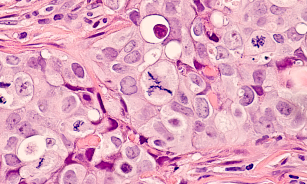
Photomicrograph of a breast cancer (grade 3 invasive ductal carcinoma) with frequent mitoses (mitotic figures), including a large central atypical mitoses.

The Trending with Impact series highlights Oncotarget publications attracting higher visibility among readers around the world online, in the news, and on social media—beyond normal readership levels. Look for future science news about the latest trending publications here, and at Oncotarget.com.

Listen to an audio version of this article

Triple-negative breast cancer (TNBC) accounts for 10-15% of all breast cancers. “Triple-negative” in this subtype of breast cancer cell refers to the lack of HER2 protein and estrogen and progesterone receptors. This means that TNBC cannot be treated with hormone inhibition and must be treated with conventional chemotherapy. In addition, many of these breast cancer cells can opportunistically switch between proliferation and quiescence—a difficult phenotype to treat. Patients diagnosed with this highly adaptable cancer frequently relapse and develop resistance to treatments.

In 2021, researchers from The University of Texas MD Anderson Cancer Center conducted a research study in hopes of developing a safe and effective therapeutic combination to treat resistant triple-negative breast cancer. Their paper, published in Oncotarget’s Volume 12, Issue 7, was entitled: “Inhibition of resistant triple-negative breast cancer cells with low-dose 6-mercaptopurine and 5-azacitidine.” 

The Study

“Evidence suggests that SUM149-metabolic adaptable (MA) cells are a suitable model of resistant human triple-negative breast cancer (TNBC) cells that can survive bottlenecks in the body, including therapeutic interventions, by opportunistically switching between quiescence and cell proliferation [578].”

In this in vitro study, researchers cultured three highly drug-resistant and metastatic progenitor-like TNBC cell lines with opportunistic switching between quiescence and proliferation. Researchers focused on designing a safe treatment that is effective in both low- and high-risk patients. The researchers note that it was critical to their study that the regimen is proven safe to administer to patients for early use in the minimal residual disease (MRD) stage after surgery, and before clinical metastasis is detected.

“For a potential therapy to be suitable at the MRD stage, it must be safe (an important criterion prior to clinical relapse) and disrupt heterogeneous progenitor-like cancer cells that evolve into clinical metastases.”

Two chemotherapy and immunosuppressive drugs (ribonucleoside analogues) were tested on the cell lines at low doses for the sake of viability in the MRD stage: 6-mercaptopurine (6-MP) and 5-azacitidine (5-AzaC). Both of these drugs have been clinically proven to be well-tolerated and to have drug-sensitizing, quiescence-stabilizing, and apoptosis-inducing effects in cancer cells.

“We chose 5-AzaC because it could complement 6-MP’s effects on the transcriptome and epigenome, and—as indicated by many Phase 1 clinical trials—5-AzaC is well tolerated [11].”

Results & Conclusion

“Our studies suggest that low-dose 6-MP, which is a purine analogue and very effective in maintaining remission in IBD [9], inhibits highly adaptable TNBC cells in our model, presumably by disrupting their transcriptome and epigenome.”

Researchers found that these low-dose therapeutics take several weeks to become effective. Despite the low dose, 6-MP (complimented by 5-AzaC) was capable of inhibiting highly adaptable TNBC cells. The researchers also point out that, based on decades of 6-MP’s use in patients with inflammatory bowel disease (IBD), this drug may be used regularly to modulate the immune system and prevent disease recurrence through its ability to inhibit chronic inflammation associated with advanced cancers.

“We suggest that low dose 6-MP and other drugs that would complement 6-MP’s action, such as 5-AzaC, could be suitable for preventing recurrence and metastasis in high-risk breast cancers. 6-MP could be taken lifelong if it is necessary for maintaining a long-term remission.”

Click here to read the full scientific study, published by Oncotarget.

YOU MAY ALSO LIKE: Latest Oncotarget Videos Hosted on LabTub TV

Oncotarget is a unique platform designed to house scientific studies in a journal format that is available for anyone to read—without a paywall making access more difficult. This means information that has the potential to benefit our societies from the inside out can be shared with friends, neighbors, colleagues, and other researchers, far and wide.

For media inquiries, please contact media@impactjournals.com.

Trending with Impact: Benign, Borderline, and Malignant Breast Tumors


Researchers conducted a 2021 study to better characterize phyllodes tumors and other breast fibroepithelial lesions in order to improve diagnosis and treatment for patients.

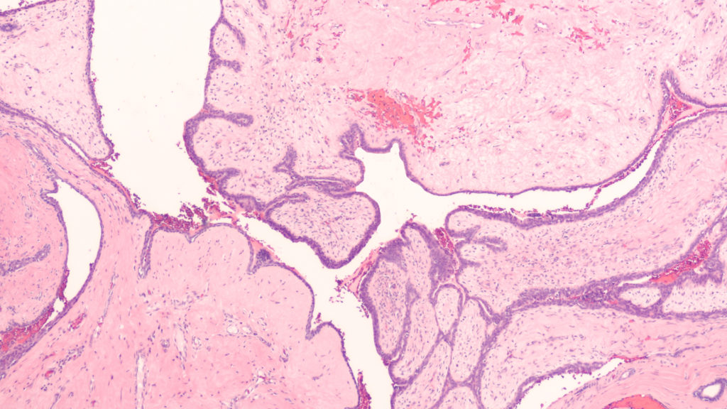
Photomicrograph showing histology of a benign phyllodes tumor of the breast, from sections of an excision specimen (lumpectomy).

The Trending with Impact series highlights Oncotarget publications attracting higher visibility among readers around the world online, in the news, and on social media—beyond normal readership levels. Look for future science news and articles about the latest trending publications here, and at Oncotarget.com.

Listen to an audio version of this article

Thankfully, around 80% of lumps found in human breasts turn out to be benign, or indolent, fibroadenoma (FAD). FADs fall into a category of breast fibroepithelial lesions (FELs), which include many heterogeneous pathological tumors, ranging from benign FADs to rare and potentially aggressive phyllodes tumors (PTs). After examination by a physician, these FELs may be diagnosed as either benign, borderline, or malignant. 

However, there is a need to improve accurate diagnosis and distinction between FELs using a marker-based diagnostic approach. In an effort to better characterize FELs, researchers from India’s CSIR-Centre for Cellular and Molecular Biology, Institute of Bioinformatics, Gandhi Hospital, Government Medical College, and Manipal Academy of Higher Education conducted a trending 2021 study, titled: “Quantitative proteome profiling stratifies fibroepithelial lesions of the breast.”

“The current grading system remains unreliable in differentiating these tumors due to histological heterogeneity and lack of appropriate markers to monitor the sudden and unpredictable malignant transformation of PTs.”

The Study

To begin identifying the differentially expressed genes and proteins among FADs and PTs in benign, borderline, and malignant states, the researchers conducted quantitative global proteomics on Formalin-Fixed Paraffin-Embedded (FFPE) tissue sections. They conducted a principal component analysis of the protein expression matrix to identify the overlapping proteomic profiles among FELs.

“Interestingly, we observed FADs and benign PTs clustered together compared to borderline and malignant ones, albeit with overlapping protein expression profiles.”

When FADs were compared with benign PTs, the researchers identified 32 proteins in FAD that were differentially regulated. The researchers elucidated many important distinctions between benign, borderline, and malignant FADs and PTs, and identified at least three potential prognostic markers that may aid in patient diagnosis and treatment. The progression of PTs from borderline to malignant and their mechanistic framework was clearly explained by the researchers in this study.

“The presence of extensive ECM proteins and EMT markers led us to hypothesize a model of deposition and degradation of these proteins thus triggering ECM remodeling and EMT acquisition in borderline PTs leading to its malignant state. Enrichment of platelet degranulation factors in malignant PT indicates active angiogenesis during this transformation.” 

The Study

“Herein, our initial findings suggest that MUCL1, HTRA1, and VEGFD can be used as potential proteomic markers that could augment existing diagnosis, and help in monitoring the progression of the disease.” 

Additional characterization of FELs using different omics platforms was recommended by the researchers to help better understand and manage the dynamics of PTs and malignant breast tumors.

“The present work shed light on a brief mechanistic framework of PTs aggressive nature and present potential biomarkers to differentiate overlapping FELs that would be of practical utility in augmenting existing diagnosis and disease management for this rare tumor.”

Click here to read the full scientific study, published in Oncotarget.

Oncotarget is a unique platform designed to house scientific studies in a journal format that is available for anyone to read—without a paywall making access more difficult. This means information that has the potential to benefit our societies from the inside out can be shared with friends, neighbors, colleagues, and other researchers, far and wide.

For media inquiries, please contact media@impactjournals.com.
Trending with Impact: New Prognostic Parameters for Breast Cancer

Different imaging and assessment tools across multiple clinics can result in varied prognostic values. Researchers from Japan conducted a retrospective study of harmonized pretreatment volume-based quantitative FDG-PET/CT parameters for prognostic values in breast cancer patients.

PET Scan image of whole body Comparision Axial, Coronal plane in patient breast cancer recurrence treatment.

The Trending with Impact series highlights Oncotarget publications attracting higher visibility among readers around the world online, in the news, and on social media—beyond normal readership levels. Look for future science news and articles about the latest trending publications here, and at Oncotarget.com.

Listen to an audio version of this article

Breast cancer consists of a wide variety of tumor types, symptoms, disease progression courses, and responses to treatments. In the clinic, researchers decide which disease interventions to use by evaluating the patients’ stage of tumor-node-metastasis (TNM), histologic tumor grade, and the levels of hormone receptors and molecular markers that are present.

Standardized uptake value (SUV), metabolic tumor volume (MTV), and tumor lesion glycolysis (TLG) are derived from 18F-fluorodeoxyglucose positron emission tomography/computed tomography (FDG-PET/CT). These variables have also been reported to correlate with clinicopathological prognostic factors and are considered predictive factors of prognosis.

Breast Cancer Prognostic Parameters

“Recently, noninvasive diagnostic tools have been gaining popularity for prediction of tumor behavior, with magnetic resonance spectroscopy (MRS) and diffusion-weighted imaging (DWI) with magnetic resonance imaging (MRI) reported to provide surrogate imaging biomarkers showing correlations with clinicopathological prognostic factors [23].”

In a multi-institutional retrospective study in Japan, researchers—from the Hyogo College of Medicine, Nippon Medical School Hospital, National Cancer Center Hospital, Kinki University Faculty of Medicine, and Gunma Prefectural College of Health Sciences—explain that the factors and algorithms used by different assessment tools across multiple clinics can result in varied standardized uptake values. These inconsistencies have provided an opportunity for the researchers to standardize parameters of prognostic values when imaging breast cancer patients to improve patient outcomes.

“Thus, a harmonization strategy is necessary for comparing semi-quantitative PET parameters among available imaging methods, which is a notably relevant issue for multicenter trials employing different PET systems.”

The Study

Researchers gathered records of 546 patients treated from 2010 to 2016 with stage I–III invasive breast cancer. Of those patients, 344 were estrogen receptor (ER)-positive/human epidermal growth factor receptor two (HER2)-negative, 110 were HER2-positive, and 92 were triple-negative. The patients were treated at four separate institutions using different PET/CT scanner systems. In addition to surgeries, chemotherapy, and radiotherapy, patients were assessed during their follow-up appointments.

“Mammography, ultrasound, CT, bone scanning, and FDG-PET/CT were used for determining disease recurrence, metastasis, and progression during follow-up.”

Researchers in this study retrospectively performed histological and statistical analyses of overall survival and recurrence-free survival in patients of each breast cancer subtype group.

“An experienced reader (12 years of experience with oncologic FDG-PET/CT) who had no knowledge of other imaging results or clinical and histopathologic data retrospectively reviewed all of the FDG-PET/CT images.”

They found that the average maximum standardized uptake values (SUVmax) for HER2-positive and triple-negative tumor patients were higher than in patients with ER-positive/HER2-negative tumors.

“Harmonized primary tumor and nodal maximum SUVmax, metabolic tumor volume (MTV), and TLG indicated in pretreatment FDG-PET/CT results were analyzed.”

Conclusion

Results from this study suggest that harmonized PET classifications with final clinical response assessments demonstrate a better ability to predict disease-free survival compared to non-harmonized PET classification.

“We concluded that harmonized quantitative volume-based values, especially those for the primary tumor and nodal SUVmax and TLG, obtained with FDG-PET/CT can provide useful information regarding prognosis for both recurrence and death in patients with operable invasive breast cancer, including all three main subtypes. The findings presented here are considered useful for improving care of individual patients.”

Click here to read the full retrospective study, published in Oncotarget.

Oncotarget is a unique platform designed to house scientific studies in a journal format that is available for anyone to read—without a paywall making access more difficult. This means information that has the potential to benefit our societies from the inside out can be shared with friends, neighbors, colleagues and other researchers, far and wide.

For media inquiries, please contact media@impactjournals.com.
Trending with Impact: Review of HER2 Variants in Breast Cancer Tumors

This review compiles splice variations in HER2 breast cancer, specifically in the context of the tumor environment, and co-expression of variants. The study also provides an up-to-date (as of Nov. 2020) account of HER2 and HER2 variant patterns of resistance to anti-HER2 therapies and other interventions.

Photomicrograph of immunohistochemistry for HER2, showing positive cell membrane staining in this infiltrating ductal carcinoma

The Trending with Impact series highlights Oncotarget publications attracting higher visibility among readers around the world online, in the news, and on social media—beyond normal readership levels. Look for future science news and articles about the latest trending publications here, and at Oncotarget.com.

According to Cancer Research UK, breast cancer occurs in one in every eight women within their lifetime and is the second highest cause of cancer related deaths in the UK. Breast cancer is a blanketed term for a wide variety of tumors that occur in the mammary glands. In over 20% of breast cancers, the human epidermal growth factor receptor two gene, officially named ErbB2 but otherwise known as HER2, is overexpressed. HER2 is a member of the epidermal growth factor receptor family of EGFR, HER2, HER3, and HER4. Overexpression of the HER2 protein was discovered in 1987 as a biomarker associated with poor prognosis and aggressive tumor types in breast cancer. This finding has accelerated research studies and progress in HER2 diagnostic testing and targeted therapeutics. However, the issue of HER2 resistance in these targeted therapies remains problematic.

“At the present time, we have an incomplete understanding of why patients with HER2+ breast cancer exhibit variable responses or resistance to targeted therapies [7374].”

Researchers from the Translational and Clinical Research Institute at Newcastle University in the United Kingdom have compiled a review of variations in HER2 breast cancer, specifically in the context of the tumor environment and when multiple variants are co-expressed at altered ratios. Their study also provides an up-to-date (as of Nov. 2020) account of the current landscape of HER2 variants and links this to patterns of resistance against HER2 therapies and other interventions.

“It is clear HER2 expression is not as simple as a single oncogenic overexpressed protein. It is likely many variants, arising from splicing and other mechanisms, are present in tandem. The relative ratios of these are likely to fluctuate depending on cellular conditions, during tumorigenesis and breast cancer progression.”

HER2 Variants & Co-expression

This paper provides an exquisitely detailed description and explanation of the HER2 protein structure, signaling pathways, sub-typing, and in-depth treatment functionality of a number of different HER2 targeted therapeutics. 

“Different forms of the HER2 protein exist within tumours in tandem and can display altered biological activities.” 

The unique interest in researching variations in HER2 breast cancer has increased since the identification of Δ16-HER2: a particular splice variant and link to resistance of anti-HER2 therapies. The “Δ16” in Δ16-HER2 refers to the lack of exon-16, which encodes a small extracellular portion of the DNA. Δ16-HER2 represents approximately 9% of the normal HER2 transcripts and its expression is considered common in breast cancer. Previous studies have identified Δ16-HER2 and HER2 normal transcripts can be co-expressed at varying levels in breast carcinomas. 

In the variant P100, less is known about this truncated HER2 protein. It has been hypothesized that P100 reduces the efficacy of monoclonal antibody HER2 treatments.

The splice variant Herstatin is produced by the retention of intron-8 in the HER2 protein. Herstatin acts as a tumor suppressor by blocking HER2 activity and cell proliferation, while promoting apoptosis. The researchers mention that it is important to note that cells expressing high levels of Herstatin are more sensitive to Tamoxifen.

“It’s noteworthy that one study proposed that the presence of Herstatin transcript does not segregate by tumour grade or size, patient age, lymph node involvement or ER status and that mRNA transcripts were present in matched non-cancerous breast tissue and breast carcinomas [96].”

Researchers in this review note that identifying and assessing the expression ratios of these different variants and classifying them as prognostic and predictive biomarkers may aid in further personalized treatment of breast cancer in HER2 positive patients.

Testing and Research Landscape

“Studying splicing regulation and how this is altered in breast cancer could explain patterns of expression and how these link to treatment resistance [111].”

The researchers write that tests assessing for both HER2 status and HER2 variant expression could potentially refine their predictions of a patients’ response to treatment. One common way that researchers gauge levels of HER2 proteins, and only some HER2 variants, is through immunohistochemistry tests. mRNA assessments are also used to identify gene expression patterns. Another biomarker test the researchers noted that may be best used for prognostic predictions is the Enzyme-Immunoassay—to assess levels of plasma or serum HER2 (sHER2) in the blood produced by cleavage or splicing.

“Cohort studies have identified sHER2 testing as a useful complementary test to IHC owing to the correlations between high sHER2 and aggressive tumour phenotypes such as invasion and metastases.”

Targeted HER2 Treatments

The review elaborates in detail about targeted treatments for HER2 breast cancer, which include: trastuzumab, pertuzumab, lapatinib, and T-DM1. They note that endocrine therapy is utilized for ER positive patients and chemotherapy, radiotherapy, and surgery are all still utilized.

Conclusion

“Work in vitro and in vivo as well as analysis from clinical trials has identified patterns of resistance to the standard of care treatment options in HER2+ patients which are correlated to variant expression.” 

This goal of their review was to summarise the current landscape of HER2 variant research and to explain why researchers should consider HER2 variant levels and ratios when offering the best treatment plan for breast cancer patients.

Click here to read the full review, published in Oncotarget.

Oncotarget is a unique platform designed to house scientific studies in a journal format that is available for anyone to read—without a paywall making access more difficult. This means information that has the potential to benefit our societies from the inside out can be shared with friends, neighbors, colleagues and other researchers, far and wide.


For media inquiries, please contact media@impactjournals.com.

Oncotarget Launches New Special Collection on Breast Cancer

Listen to an audio version of this post

As you may know, Oncotarget is a scientific journal that publishes oncology-focused review and research papers every week on its open access platform — available at no cost to readers. Recently, a new Special Collections series debuted, and the first collection launched in honor of breast cancer awareness.

What makes our collections special? 

Oncotarget carefully selects the most credible and insightful studies to publish on Oncotarget.com, while also choosing papers that link different fields of oncology, cancer research, and biomedical sciences together to eliminate borders between specialties. The term “oncotarget” encompasses all molecules, pathways, cellular functions, cell types, and tissues that can be viewed as targets relevant to cancer, as well as other diseases. This journal is a resource for oncology researchers and the larger scientific community.

Before a study is published in Oncotarget, selected papers are meticulously peer-reviewed by an editorial board of award-winning scientific editors from academic universities and institutions well-known for their excellence and precision. Click here for a complete list of Oncotarget Editorial Board members.

Breast cancer research

Each year, over 40,000 women and men lose the fight against breast cancer in the United States. After skin cancer, breast cancer is the most commonly diagnosed cancer in women. The spread of breast cancer awareness and increase in research funding has helped develop advances and discoveries in the diagnosis and treatment of this proliferous cancer. 

The new Special Collections by Oncotarget are yet another tool researchers and science readers alike may use as a resource to learn more about breast cancer. The creators of these collections also hope that they may be used by scientists to discover new biomarkers, mechanisms, and therapies to improve our quality of life and better treat cancer and diseases.

Click here to explore the Special Collection on breast cancer.

Thanks to Impact Journals, we know exercise helps to fight breast cancer—for free

oncotarget

A recent breakthrough medical study has revealed that exercise has been proven to combat breast cancer.  The paper, entitled “Anticancer effect of physical activity is mediated by modulation of extracellular microRNA in blood,” was recently published in a June 2020 issue of the free online open-access medical journal Oncotarget. It was authored by an international team of medical researchers, headed by Dr. Alessandra Pulliero of the University of Genoa in Italy, and included Doctors Ming You, Pradeep Chaluvally-Raghavan, Barbara Marengo, Cinzia Domenicotti, Barbara Banelli, Paolo Degan, Luigi Molfetta, Fabio Gianiorio, and Alberto Izzotti.

The paper has already received widespread acclaim and coverage, reproduced online by prestigious organizations such as the National Center for Biotechnology Information (a branch of the U.S. National Institutes of Health), the American Association for the Advancement of Science, and ResearchGate.

THE STUDY

While previous medical studies have shown that physical activity reduces the risk of cancer, particularly breast cancer, it’s been a mystery up to now exactly how this happens. Medical researchers have long suspected that this healing process is triggered by microRNAs, cellular fragments of RNA (ribonucleic acid) also known as miRNAs.

What’s RNA? Like DNA (deoxyribonucleic acid), RNA is one of the building blocks of life. It acts as a messenger transmitting instructions that control the synthesis of proteins. MicroRNAs stop a particular protein from being produced by binding to, and then destroying, the messenger RNA that would have produced this protein.

It is known that miRNAs are incredibly important when it comes to carcinogenesis (the creation of cancer) and cancer outcomes. In addition, MiRNAs regulate the creation of muscle tissueand muscle mass, and it’s been learned that structured exercise controls the creation of miRNA, especially in skeletal muscle.

The research team endeavored to test how exercise in breast cancer patients changed the production of miRNA in their bodies. To begin, 30 women from northern Italy between 54 and 78 years old walked for 45 minutes on the treadmill under identical conditions. Blood samples were taken from them both before and after the exercise sessions.

THE RESULTS

A technique known as microarray analysis revealed that structured exercise modified 14 different extracellular miRNAs related to cancer. Structured exercise caused all these miRNAs to decrease, except for a miRNA called miR-206, which increased. The researchers discovered that the most striking effects induced by exercise were changes in two miRNAs involved in breast cancer progression.

When the researchers investigated the biological effects of these two miRNAs on human breast cancer cells, they conclusively learned that working together, the changes in these two microRNAs activated by a physical exercise program suppressed breast cancer cells. Since too many miRNAs are linked to triggering inflammation and the creation of lymphocytes (white blood cells in the lymph system, which can influence breast cancer), the researchers also believe that structured exercise might reduce inflammation by modulating miRNA in the blood.

They also found that structured exercise improved blood pressure and glucose levels (cancerous tumors feed on glucose) among participants. The doctors discovered that these improvements in blood pressure and glucose levels helped regulate the miRNAs being studied, and in turn helped the miRNAs combat cancer.

This international team of researchers is confident that by testing for the levels of these miRNAs in patients’ blood, they’ve achieved a non-invasive way of establishing biomarkers (a measurable sign of whether a disease is present or how severe it is) to prevent breast cancer. This is potentially a significant breakthrough in breast cancer prevention and treatment.

As a result of this study, the medical community now knows that structured exercise fights breast cancer, and it’s been given a non-invasive way to diagnose and battle breast cancer—and possibly other forms of cancers as well.

ABOUT ONCOTARGET

This important study was able to be published, and noticed so quickly, because it was made available by Impact Journals’ free, open-access cancer research journal Oncotarget. Currently, over 20,000 Oncotarget papers are also searchable on PubMed, a widely used free search engine for life sciences and biomedical research. 

Because Oncotarget is open-access, it is free for everyone in the world to read. Most medical journals charge authors for publishing their work, and then in turn charge readers to access what could be all-important, life-saving information. With its revolutionary publishing model, Impact Journals, through publications like Oncotarget, makes it easy for anyone with important medical discoveries to communicate them to the public in the fastest and most effective way possible—possibly saving, prolonging, and improving many people’s lives in the process.

With the goal of a life without disease, Impact Journals allows scientists to share their exceptional discoveries, offers services that enable rapid dissemination of results, and presents vital findings from the many fields of biomedical science. It shares scientific findings through a comprehensive publication process entailing peer review, manuscript preparation, and publication promotion.

In addition, Oncotarget is well-known for publishing papers by Nobel Prize winners. The 2019 Nobel Prize in Physiology or Medicine was awarded jointly to Oncotarget Editorial Board members William G. Kaelin Jr., and Gregg L. Semenza for their discoveries of “how cells sense and adapt to oxygen availability,” which can help us understand and potentially treat a range of conditions like cancer, heart attack, stroke, and anemia. (They shared the Prize with UK physician-scientist Sir Peter J. Ratcliffe.) Both William G. Kaelin and Gregg L. Semenza are founding members of Oncotarget, where Gregg L. Semenza has published eight papers.

Another notable Oncotarget Nobel Prize winner is endocrinologist Andrew V. Schally, a member of Oncotarget’s Editorial Board who won the Nobel Prize in Physiology or Medicine in 1977 and who has published 12 papers in Oncotarget. Of Oncotarget’s work, he remarked: “Oncotarget is an outstanding and most important journal in the field of oncology and cancer research. Oncotarget is performing an extremely useful function for those of us working not only in cancer research, but also on other important topics in the field of medicine. Oncotarget deserves strong support from investigators working in the area of oncology as well as from the National Institutes of Health (NIH).”

If you would like to be first to learn about some of the most exciting new discoveries in medical science, consider investigating the groundbreaking work being published by Impact Journals, including its flagship publication, Oncotarget.